- This topic is empty.
-
AuthorPosts
-
In the realm of analytical chemistry, the detection and quantification of ferrous ions (Fe²⁺) in various samples, particularly water, is of significant importance. Ferrous ions play a crucial role in numerous environmental and industrial processes, and their accurate measurement is essential for water quality monitoring, industrial effluent control, and research applications. In this blog post, METASH, a high performance laboratory analysis equipment exporter, will share the detection of ferrous ions using double beam uv-visible spectrophotometer for sale.
Introduction
The traditional method for detecting ferrous ions often involves the use of the o-phenanthroline spectrophotometric method, which is standardized in protocols such as HJ/T 345-2007 for water quality analysis. This method typically requires a heating step to ensure the complete reduction of ferric ions (Fe³⁺) to ferrous ions (Fe²⁺), which then react with o-phenanthroline to form a colored complex that can be measured at a specific wavelength. However, this heating step has been identified as a major bottleneck in the process, consuming a significant amount of time and often leading to inconsistencies due to uneven heating. The recent technical advancement discussed in this article aims to address these challenges by eliminating the heating step and optimizing the use of reagents, thereby reducing the overall experimental time while maintaining the precision and accuracy of the method.
Experimental Methodology
Instruments and Reagents
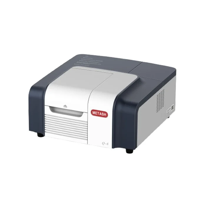
The primary instrument utilized in this study is the Q-8 double beam UV-visible spectrophotometer, which offers high precision and reliability for quantitative analysis. Other essential equipment includes a heating plate, an analytical balance with a precision of 0.0001 g, a pipettor, and a funnel. The reagents required for this method include hydroxylamine hydrochloride, glacial acetic acid, ammonium acetate, sodium acetate, o-phenanthroline, ammonium iron(II) sulfate, and hydrochloric acid.

Preparation of Standard Solutions
1. Standard Reserve Solution: Accurately weigh 0.1755 g of ammonium iron(II) sulfate ((NH₄)₂Fe(SO₄)₂·6H₂O) and dissolve it in 50 mL of sulfuric acid solution (1:1 v/v). Transfer the solution to a 250 mL volumetric flask and dilute to the mark with water. This solution contains 100 µg of iron per milliliter.
2. Standard Working Solution: Pipette 25.0 mL of the standard reserve solution into a 100 mL volumetric flask and dilute to the mark with water. This working solution contains 25.0 µg of iron per milliliter.
Preparation of Reagents
1. O-Phenanthroline Solution (0.5%): Weigh 0.5 g of o-phenanthroline, dissolve it in a minimal amount of ultra-pure water, add a few drops of hydrochloric acid, and then dilute to 100 mL.
2. Buffer Solution: Weigh 40.0 g of ammonium acetate, dissolve it in 50 mL of glacial acetic acid, and then dilute to 100 mL.
3. Reducing Agent (10% Hydroxylamine Hydrochloride): Weigh 10.0 g of hydroxylamine hydrochloride, dissolve it in a minimal amount of ultra-pure water, and then dilute to 100 mL.
4. Saturated Sodium Acetate Solution: In a 100 mL beaker, add 50 mL of ultra-pure water and dissolve sodium acetate until no more can be dissolved.
Standard Curve Construction
Two approaches were employed to construct the standard curve: the traditional heating method and the simplified non-heating method.
1. Heating Method: Pipette 0, 2.0, 4.0, 6.0, 8.0, and 10.0 mL of the standard working solution into 250 mL Erlenmeyer flasks, respectively. Add distilled water to make up to 50.0 mL, followed by 1 mL of hydrochloric acid solution (1:3 v/v) and 1 mL of 10% hydroxylamine hydrochloride. Add 1–2 glass beads and heat to boil until the solution is reduced to approximately 15 mL. Cool to room temperature and transfer quantitatively to 50 mL stoppered colorimetric tubes. Add a small piece of Congo red paper and titrate with saturated sodium acetate solution until the paper just turns red. Then add 5 mL of buffer solution and 2 mL of 0.5% o-phenanthroline solution, dilute to the mark with water, and shake well. After 15 minutes of color development, measure the absorbance at 510 nm using water as the reference.
2. Non-Heating Method: Pipette 0, 2.0, 4.0, 6.0, 8.0, and 10.0 mL of the standard working solution into 50 mL stoppered colorimetric tubes, respectively. Add 5 mL of 10% hydroxylamine hydrochloride solution to each tube, shake well, and let stand for 2 minutes. Then add 5 mL of ammonium acetate solution and 5 mL of 0.5% o-phenanthroline solution, and dilute to the mark with ultra-pure water. Allow the mixture to develop color for 15 minutes before measurement.
Sample Analysis
For the analysis of water samples, 5 mL of the sample is directly mixed with 5 mL of buffer solution and 2 mL of 0.5% o-phenanthroline solution, diluted to 50 mL, and allowed to develop color for 5–10 minutes. The absorbance is then measured at 510 nm using the reagent blank as the reference.
Results and Discussion
Standard Curve
The standard curve was constructed by plotting the absorbance values against the concentration of ferrous ions. The results showed that the simplified non-heating method produced a standard curve that was comparable to that obtained using the traditional heating method. The linear relationship between absorbance and ferrous ion concentration was maintained, indicating that the non-heating method did not compromise the accuracy of the measurements. The correlation coefficient (R²) for the standard curve was close to 1, demonstrating a strong linear relationship and the reliability of the method.
Sample Analysis
The accuracy and precision of the simplified method were further validated through the analysis of water samples. The results of the sample analysis were consistent with the expected values, and the method demonstrated good reproducibility. The recovery rates of ferrous ions in spiked samples were within the acceptable range, confirming the reliability of the method for quantitative analysis. The simplified method not only reduced the experimental time significantly but also eliminated the potential errors associated with the heating step.
Conclusion
The simplified method for detecting ferrous ions using a double beam UV-visible spectrophotometer, as described in this article, offers a significant improvement over the traditional method. By eliminating the heating step and optimizing the use of reagents, the method achieves a substantial reduction in experimental time without sacrificing precision or accuracy. This innovative approach provides a more efficient and reliable alternative for the detection of ferrous ions in water samples, making it a valuable tool for environmental monitoring, industrial applications, and research. Future work may focus on further optimizing the method and exploring its potential applications in other types of samples.
http://www.metashcorp.com
METASH -
AuthorPosts